Histology: The Basics
Start at Square One
What is Histology?
Before we jump in with both feet, let’s take a step back… what is histology anyway??
Histology is a sub-discipline of anatomy that is concerned with the microscopic structure of tissues. A tissue is an aggregation of similar cells from a common embryonic origin that work together to perform a specific collective function. There are 4 general categories of tissues in the human body (epithelial, connective, muscle, and nervous), each of which will be explored in depth on individual pages of this site!

Does this sound familiar??
Chances are it’s because you are trying to memorize everything instead of actually understanding the fundamentals… I know it sounds like a lot of work, but nothing worth having comes easy. Better yet, it will save you SO MANY sleepless nights trying to cram before your next histo exam!
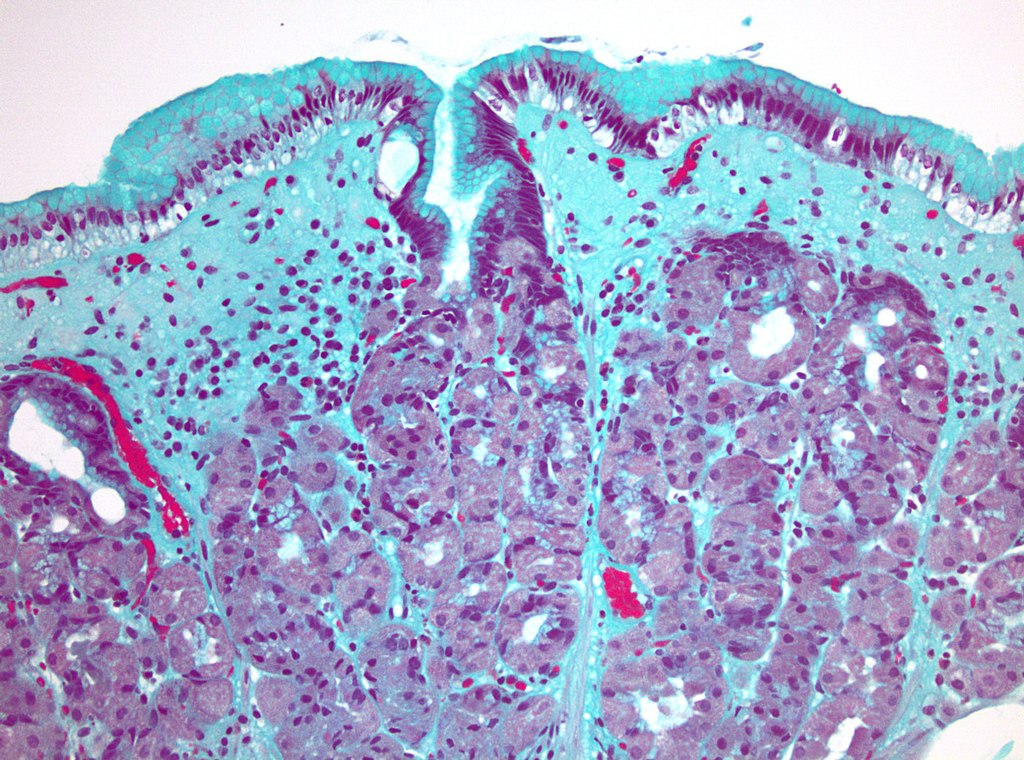
Histological Staining
Stains—including coloured dyes, labelled antibodies, and heavy metals—are used in histology to visualize and distinguish between tissue components. At baseline, most tissue components have nearly indistinguishable optical densities. Histological stains that bind to specific components and/or bind to different extents can be used, depending on the desired outcome. Here are a few common staining techniques & a brief overview of how they work:
1) Haematoxylin and Eosin (H&E):
A routine way to differentiate between tissue components is with a combination of acidic and basic dyes. A basic dye has a positive net charge and will bind to negatively-charged components. In contrast, an acidic dye has a negative net charge and will bind to positively-charged components. One of the most common combinations is haematoxylin (basic dye: stains purple/blue) & eosin (acidic dye: stains pink/red).
Quick throwback to high school biology:
- Acidic structures (-) are basophilic & will therefore be stained by basic dyes (+)
- Basic structures (+) are acidophilic & will therefore be stained by acidic dyes (-)
ACIDIC (Basophilic) STRUCTURES
[Basic Dye: Purple/Blue]
DNA & RNA (e.g. nucleus / nucleolus)
RNA (e.g. ribosomes /
rough endoplasmic reticulum)
BASIC (Acidophilic) STRUCTURES
[Acidic Dye: Pink/Red]
Basic proteins (e.g. in cytoplasm)
Collagen (e.g. connective tissues)
Red Blood Cells
*Note: H&E staining (like all histological staining) can be influenced by numerous factors, including tissue preparation, staining protocol, and source & batch of stain (e.g. pH of stain).

2) Masson’s Trichrome:
As the name suggests, Masson’s trichrome (and any other trichome stains) involves 3 colours of dye. This particular trichrome is useful for distinguishing muscle fibres from collagen fibres!
- Tissues are fixed in an acid-resistant basic dye (Weigert’s iron Haematoxylin) which will stain acidic structures (i.e. nuclei & other RNA/DNA-containing structures) purple/blue
- An acidic plasma dye (e.g. Biebrich scarlet-acid fuchsin) will then dye all basic (acidophilic) structures pink/red
- Collagen is then decolourized with phosphomolybdic / phosphotungstic acid and then dyed green/blue with a collagen fibre stain (e.g. light green or aniline blue)
Basic Dye: Purple/Blue
RNA/DNA-containing structures (e.g. nuclei)
Acidic Dye: Pink/Red
Muscle fibres
Cytoplasm
Acidic Dye: Blue/Green
Collagen
Bone
3) Nissl Stains:
Nissl bodies (or Nissl substance) specifically refers to granular endoplasmic reticulum and ribosomes found primarily in neuronal cell bodies and dendrites. Nissl stains—like cresyl violet or toluidine blue—are basic dyes that will bind with the negatively-charged (basophilic) nucleic acids found in DNA & RNA, staining them purple or blue (depending on the chosen stain). Nissl stains will non-selectively stain all types of neurons in brain or spinal cord sections, but will not reveal any detailed information about neuronal morphology (like Golgi stains).

Histology vs. histochemistry vs. immunohistochemistry??
Turns out there are many terms that fall under the umbrella of “histology”!
I am definitely guilty of using these 3 terms interchangeably (oops!), so here’s what I’ve gleaned so far about how they differ:
Histology
The overarching study of the microscopic anatomy of biological tissues
Histochemistry
Specific branch of histological staining concerned with the chemical composition of biological tissues at a microscopic level
Immunohistochemistry*
Specialized branch of histological staining that uses selectively-binding antibodies to target specific antigens within biological tissues
* admittedly my default term when I’m trying to sound fancy
Microscopy
Since we’ve established that histology is concerned with microscopic tissue structure, it goes without saying that some magnification is essential! There are numerous types of microscopy, but light microscopy (also called optical microscopy) is one of the most common. Let’s take a closer look…

Light Source: As the name light microscopy implies, a light source is equipped to the base of the microscope to illuminate the specimen.
Condenser: Located above the light source, the condenser collects and focuses light on the specimen through a hole (called the aperture) in the stage. In many light microscopes there is also a diaphragm (or iris) located between the condenser and the stage to regulate the amount/intensity of light that reaches the specimen.
Slide: The star of the show ★—a slide with a sectioned & stained tissue sample—is placed on the aptly named stage. Clips may be present under which to secure the slide in place.
Focus Adjustment: Simply put, the coarse and fine adjustment knobs help to focus the microscope. Typically these adjustments move the stage toward or away from the objective lens.
Objective Lenses: Most microscopes have a revolving nosepiece that holds several (typically 3–4) objective lenses of different magnification powers (e.g. from 4X–100X). Objectives tend to be the most optically complex components of a light microscope, often made up of numerous internal lenses. For the sake of simplicity, all of the lens are represented as a single biconvex lens in the microscopy optics diagram below.
Eyepiece (Ocular Lens): In the immortal words of Drake, “Started from the bottom, now we’re here!” The eyepiece, or ocular lens, is the part you look through at the top of the microscope. In contrast to the interchangeable objective lenses, the eyepiece has a set magnification (usually 10X).
Total Magnification =
Objective Lens Magnification
X
Ocular Lens Magnification
NOTE: Microscopy optics may cause flashbacks to high school physics…
proceed with caution!

Recommended Resources
Chapman Histology with Dr. Jamie Chapman [YouTube]
I highly recommend starting with the “3 Minute Histology – Introduction to Histology” playlist for an overview of H&E staining and histological artefacts in < 30 minutes total!
I Heart Histo [Blog]
You’re going to love this visually-stunning, highly educational blog by Dr. Nathan Swailes! Get your weekly dose of histology, with everything from “The Histopathologist’s H&E Palette” to “Guess Who? Histology Edition”.
Staining Techniques & Troubleshooting with Dr. Damien Harkin [YouTube]
Itching to try your hand at histological staining? Dr. Harkin has you covered! Still not convinced? Look no further than the Histological Techniques Trailer!
Histology & Histopathology: Virtual Microscope [Interactive Tool]
No access to a lab? No problem! The OpenScience Laboratory (an initiative of The Open University and The Wolfram Foundation) offers several different “virtual microscope” experiments, including 320 annotated slides in the Histology & Histopathology collection!